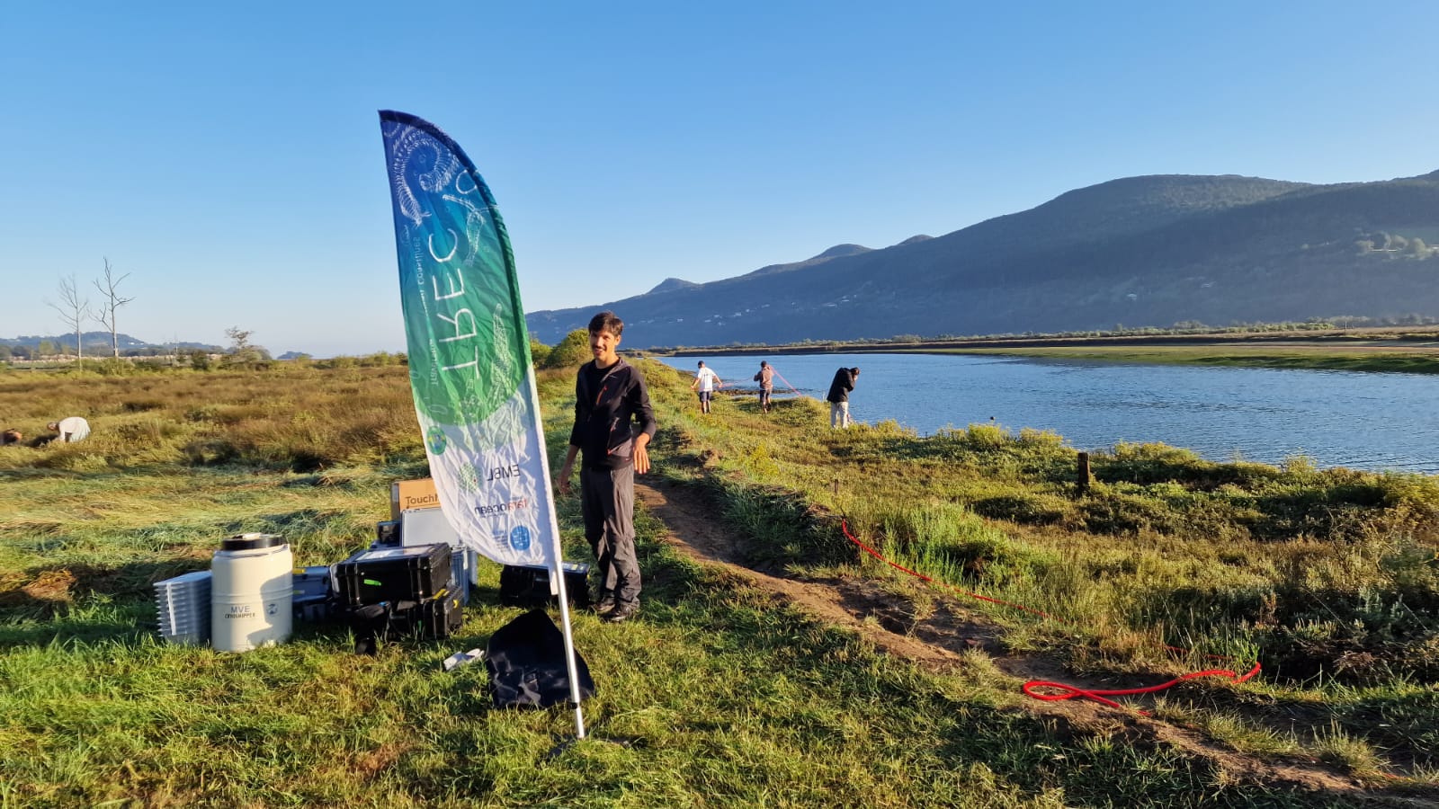
Author: Ibon Cancio, UPV/EHU Associated Professor in Cell Biology; researcher in the ‘Cell Biology & Environmental Toxicology’ (CBET) research group of the Plentzia Marine Station (PiE-UPV/EHU); Spanish scientific representative in the EMBRC Committee of Nodes

Osamu Shimomura was born in Fukuchiyama, Kyoto prefecture, Japan, on 27 August 1928. He was in school around Nagasaki in 1944, at the peak of the war’s escalade of the Japanese empire. All populations were mobilised to feed the empire’s war machine including Shimomura and his fellow students. Only 16 years old, he spent his entire 10th grade working in a Naval Aircraft Arsenal near Isayaha. There, Shimomura survived full destruction in an air raid, running among falling incendiary bombs. The fate of some of his classmates who found refuge in the bunker of the factory was worse. Graduating from school in 1945 without having attended any classes, he remained in the naval forces and was sent to the hills between Isayaha and Nagasaki to work on the repair of fighter engines. On 9 August 1945, Shimomura saw with his own eyes the B-29 plane that threw the ‘Fat Man’ and destroyed Nagasaki. In his autobiography for the Nobel Foundation we can read his account of the event:

'I went to the top of a nearby hill with a couple of friends and looked at the sky. We saw a single B-29 going from north to south towards Nagasaki, about 15 km away”… “We returned to our factory building. At the moment I sat down on my work stool, a powerful flash of light came through the small windows. We were blinded for about 30 seconds. Then, about 40 seconds after the flash, a loud sound and sudden change of air pressure followed. We were sure there was a huge explosion somewhere, but we didn’t know where. The sky was rapidly filling with dark clouds, and when I left the factory to walk home, about three miles away, a drizzling rain started. It was black rain. By the time I arrived home, my white shirt had turned gray. My grandmother quickly readied a bath for me. That bath might have saved me from the ill effects of the strong radiation that presumably existed in the black rain'.


Emperor Hirohito would announce surrender a few days later on the 15th of August and, with that, Shimomura initiated his outstanding research career in the search of light (bioluminescence). He enrolled at Nagasaki College of Pharmacy, graduating in 1951 to then work as a research student at Nagoya University. In one year, Shimomura succeeded in what others had attempted for over 20 years: purifying and crystallizing luciferin from Cypridina, a glowing crustacean common in shallow coastal waters of Japan. Luciferin (Lucifer=the light bearer) is a small molecule that, with oxygen and in the presence of an enzyme (luciferase or a photoprotein), is responsible for light emission in most bioluminescent organisms. The luciferase luciferin system was discovered in the late 19th century at a marine station in the French Mediterranean by Raphael Dubois. But this will be another history! Shimomura’s achievement opened the doors for a research opportunity in the USA thanks to an invitation from Frank H. Johnson of Princeton University in 1959. Prof Hirata decided to award Shimomura a PhD in organic chemistry for his work with Cypridina. That would allow a better salary in the USA!

At Princeton, during his first year, he continued work on available Cypridina dried samples, extracting luciferase this time. Johnson then proposed to study bioluminescence in the hydrozoan jellyfish Aequorea victoria, which was widely available on the west coast of the USA. Shimomura of course accepted. Curiously enough, Aequorea is a hydrozoan, the favourite group of creatures of yet another Japanese illustrious marine biologist: Emperor Hirohito himself. So, Shimomura, his wife Akemi, Johnson, and an assistant drove a car for seven days from Princeton to Friday Harbour in Puget Sound, Washington, in 1961. As jellyfish were continuously available there during the summer, they were available to harvest thousands of individuals per day. They were all dissected in the Friday Harbour Laboratory (University of Washington) and back at Princeton, Shimomura isolated a bioluminescent substance that turned out to be a protein. It was baptised as aequorin, the first photoprotein to be isolated. It emits blue light in the presence of calcium. Not only that, in the column chromatographies another protein co-eluted with aequorin, but this one fluorescing in green, the green fluorescent protein (GFP). Aequorin and GFP form a coupled system where aequorin produces and emits blue light that excites GFP, which in turn emits green light. Shimomura would elucidate the chromophore of GFP in 1979.

Map of Shimomura's travels across the US
Google Maps says that crossing the USA today from Princeton to Puget Sound takes 46 hours (by car). Shimomura,'s first trip, with his mentor Prof Johnson driving the whole time, took 7 days.

He then spent two years back in Japan, with a stint in New Zealand, before going back to Princeton. He then undertook yearly summer migrations to Friday Harbour to fish more jellyfish and harvest their aequorin. All together, he made 19 trips, 13 of them by car, from 1961 to 1988, in which he estimated to have collected 850,000 individuals of Aequorea. With the procedures that Shimomura could employ, it was necessary to collect around 10,000 jellyfish (0.5 tons) in order to produce 5 mg of highly purified aequorin. The consequence of hard work was determination of the structure of its luciferin, named coelenterazine. In the meantime, Ca2+ levels were discovered to be very important for different cell processes, for instance muscle contraction. Therefore, aequorin was widely used as an excellent probe to analyse cell Ca2+concentrations. Shimomura was the only source of aequorin and he sent samples to any researcher worldwide requesting them. The application of such supplies of natural aequorin reached its maximum in 1985, when a cDNA clone was produced and also additional fluorescent Ca2+ probes were made available. In 1988 Shimomura produced various semi-synthetic aequorins that had various calcium sensitivities and properties.

The wonder of hydrozoans
The wonder of hydrozoans and their 'lights'. Photo by Ibon Cancio.

In 1994, Martin Chalfie (Columbia University) introduced and expressed GFP in E. coli and in Caenorabdhitis elegans (a nematode worm), observing its fluorescence in vivo. The results suggested that GFP could be expressed in other organisms to observe the behaviour of different cells along development or under diverse physiological or environmental circumstances. Roger Tsien (University of California, San Diego), engineered GFP by modifying the amino acid residues surrounding the chromophore, producing many different fluorescent proteins emitting all the colours of the rainbow. Today, GFP is widely used to tag different proteins and molecules fluorescently, produce transgenic organisms or gene reporter assay, and it is an essential tool in the study of biology, physiology, and medicine. The applications of these fluorescent proteins coming from the oceans has brought a revolution to 21st century biomedicine.

When Shimomura’s mentor at Princeton retired, Shimomura decided that he wanted to work at a marine biological station. This decision led him to the Marine Biological Laboratory (MBL) in Woods Hole, in 1981. He worked there until his retirement as a Senior Scientist, accompanied by his wife, who worked as his research assistant. The song may say, ‘Love is in the air’, but marine biology has shown us that ‘bioluminescence is in the sea’. Shimomura studied bioluminescence and luciferin/luciferase in the limpet Latia; the krill Meganyctiphanes; the worm Chaetopterus; the firefly squid Watasenia, various coelenterates, dinoflagellates, luminous scale worms, the millipede Luminodesmus; the brittle star Ophiopsila; and several types of luminous mushrooms and bacteria.

Ascidian Phallusia mammillata embryo injected at the 16 cell stage in the A5.2 blastomere with an mRNA coding for MAP7 attached to GFP and fluorescing in green
Ascidian Phallusia mammillata embryo injected at the 16 cell stage in the A5.2 blastomere with an mRNA coding for MAP7 attached to GFP and fluorescing in green. Photo by R. Dumollard, L'institut de la Mer de Villefranche (IMEV) and deposited in movincell.org, a product of EMBRC France.


In 2008 Shimomura received the Order of Culture, the highest honour given annually by the Emperor of Japan, at a ceremony at MBL in 2008. He was elected to the U.S. National Academy of Sciences in 2013. Sorry, I was about to forget it….and the Nobel prize in Chemistry in 2008, together with Chalfie and Tsien!! He died in 2018. This was in Nagasaki, where the quest for the ‘glow’ had begun.

San Diego beach
A San Diego beach scene drawn with an eight-colour palette of bacterial colonies expressing fluorescent proteins derived from GFP and the red-fluorescent coral protein dsRed. The colors include BFP, mTFP1, Emerald, Citrine, mOrange, mApple, mCherry and mGrape. Artwork by Nathan Shaner, photography by Paul Steinbach, created in the lab of Roger Tsien in 2006 (source: Wiki Commons).


Find out more

See all news
NEWS
Discover marine biology through our PhD-focused course on marine invertebrates and live imaging. Theory meets practice with online modules and onsite labs. Led by IMEV experts, master sample prep, microscopy, and image analysis.
NEWS
For the women and girls aspiring to develop a career in STEM, it can be hard to know how to have a positive impact in science, especially without seeing strong role models. So we wanted to introduce a few of our team members and talk to them about the vital role women play in the advancement of marine science.