Press release
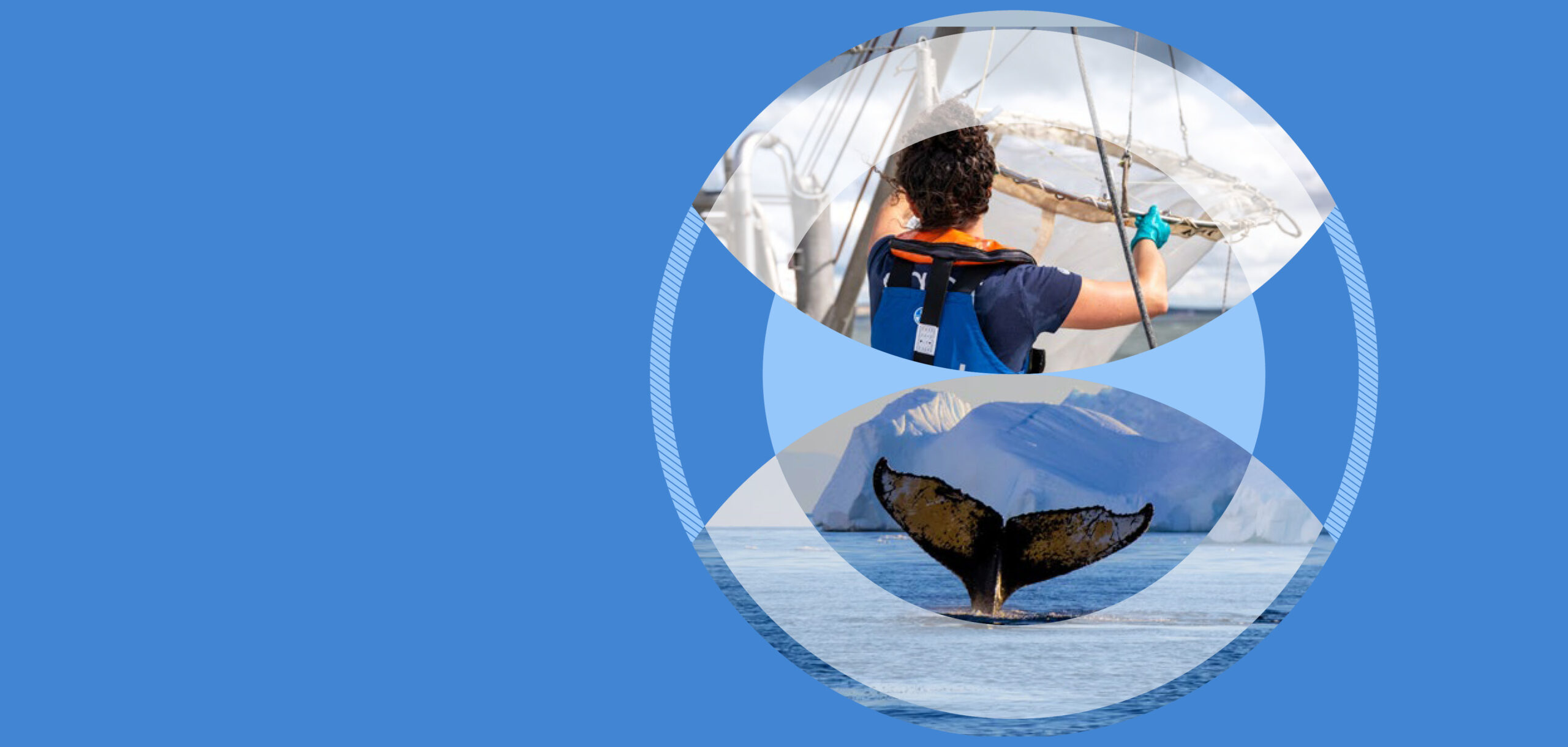
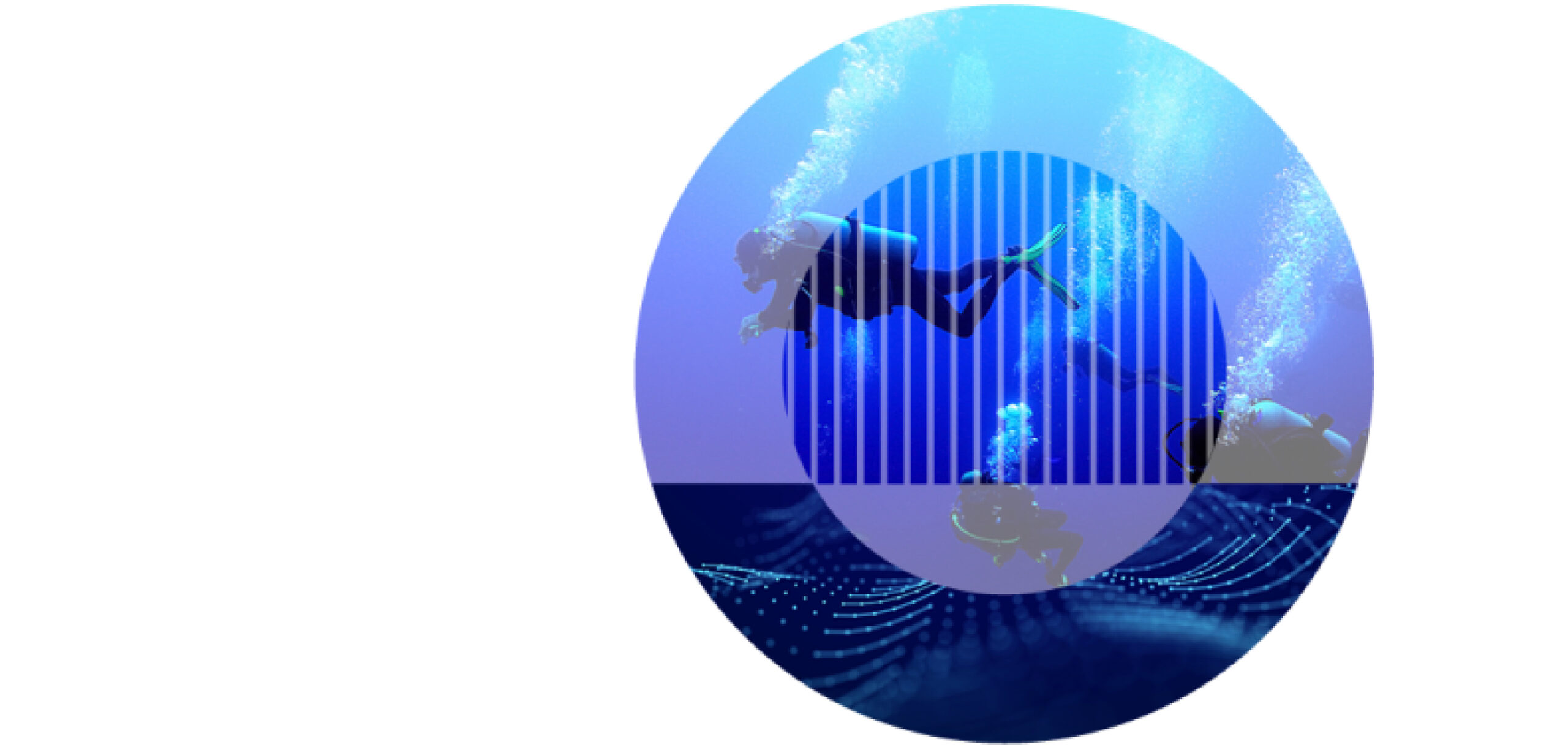
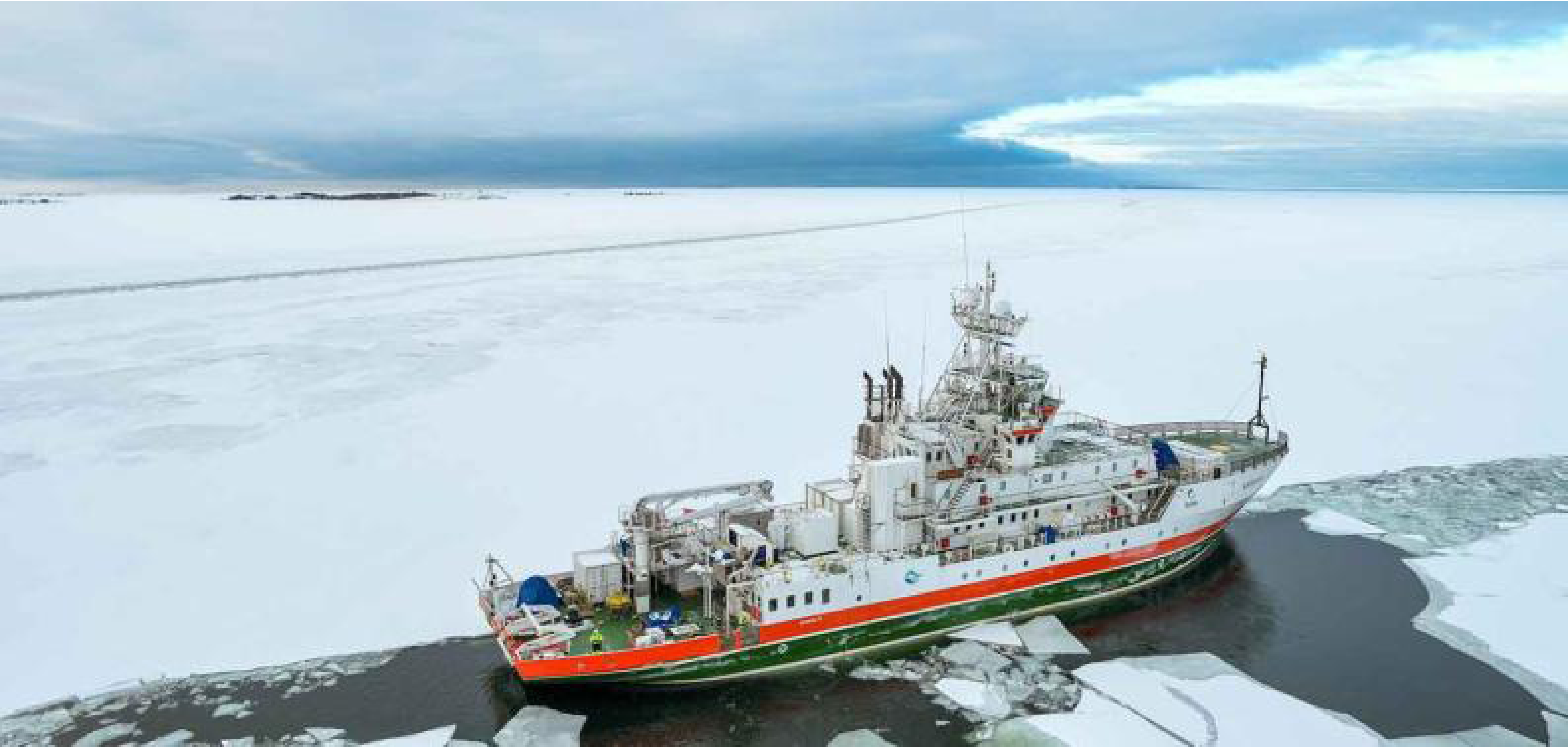
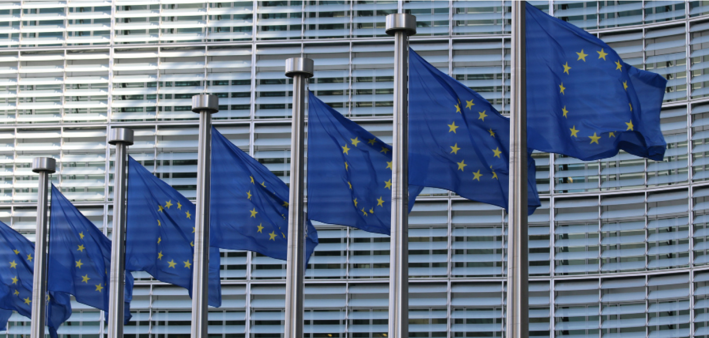
EMBL, Tara and EMBRC on an expedition to study human impact on European coastal ecosystems
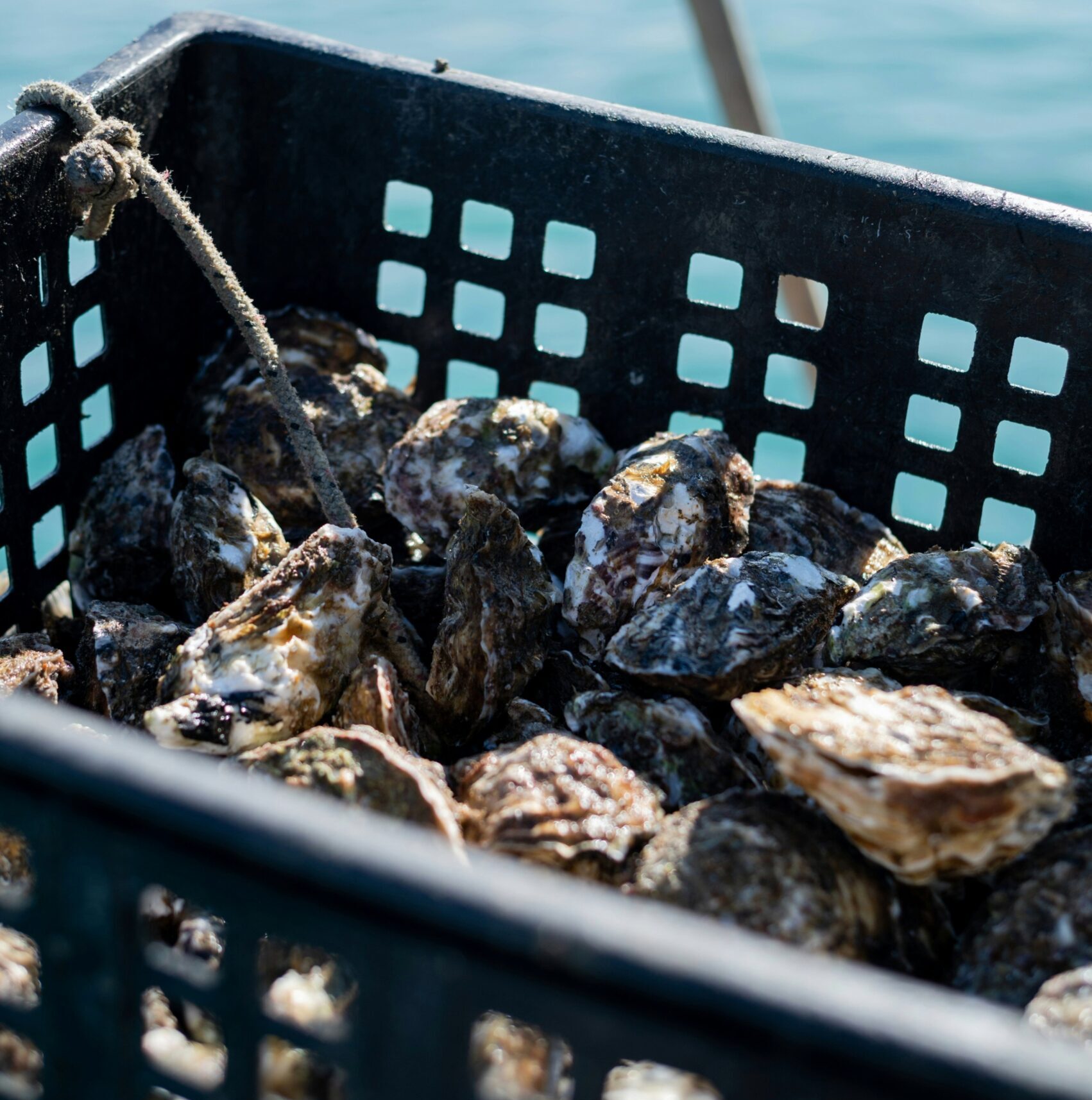
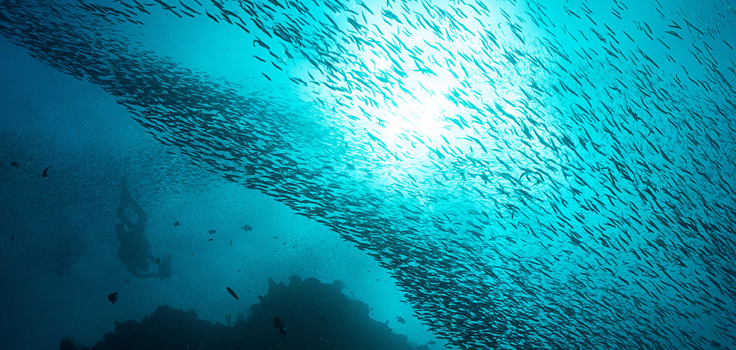
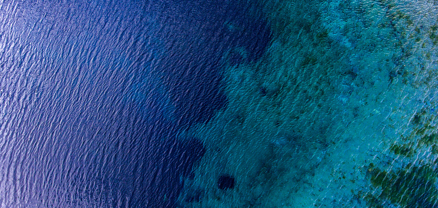
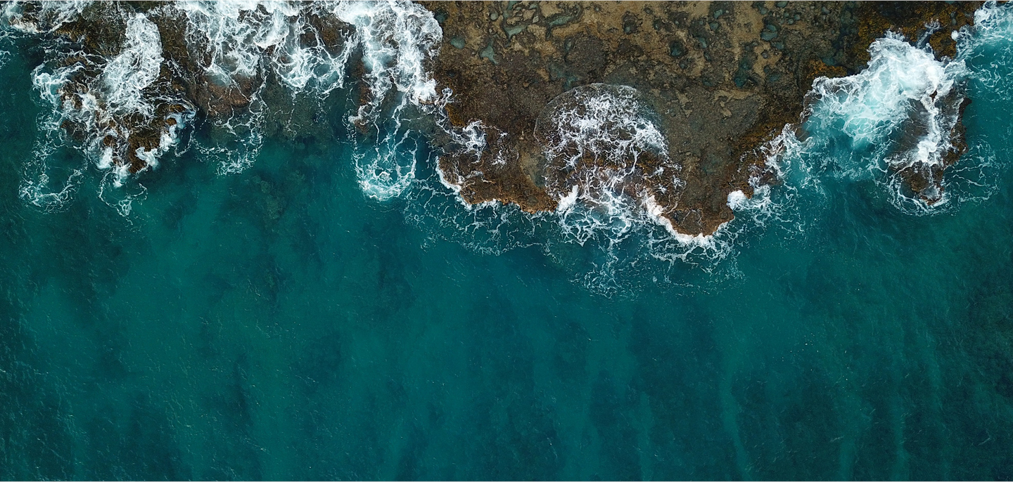
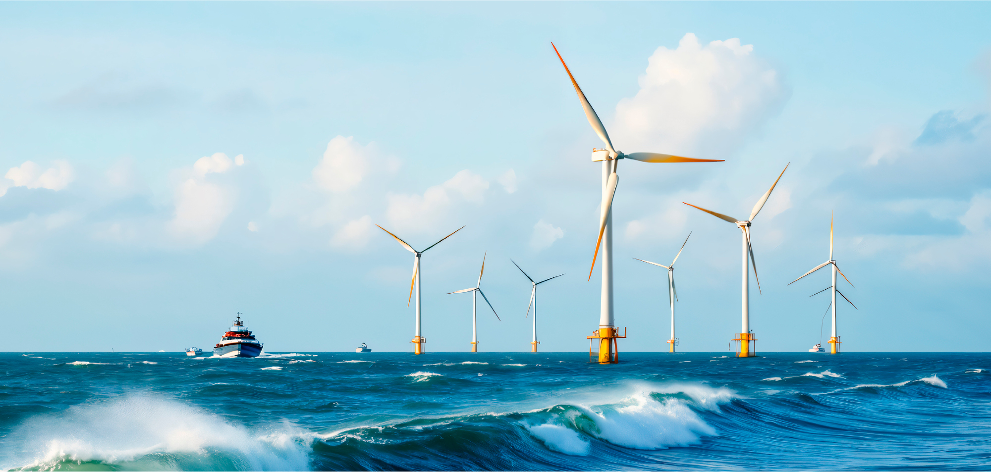
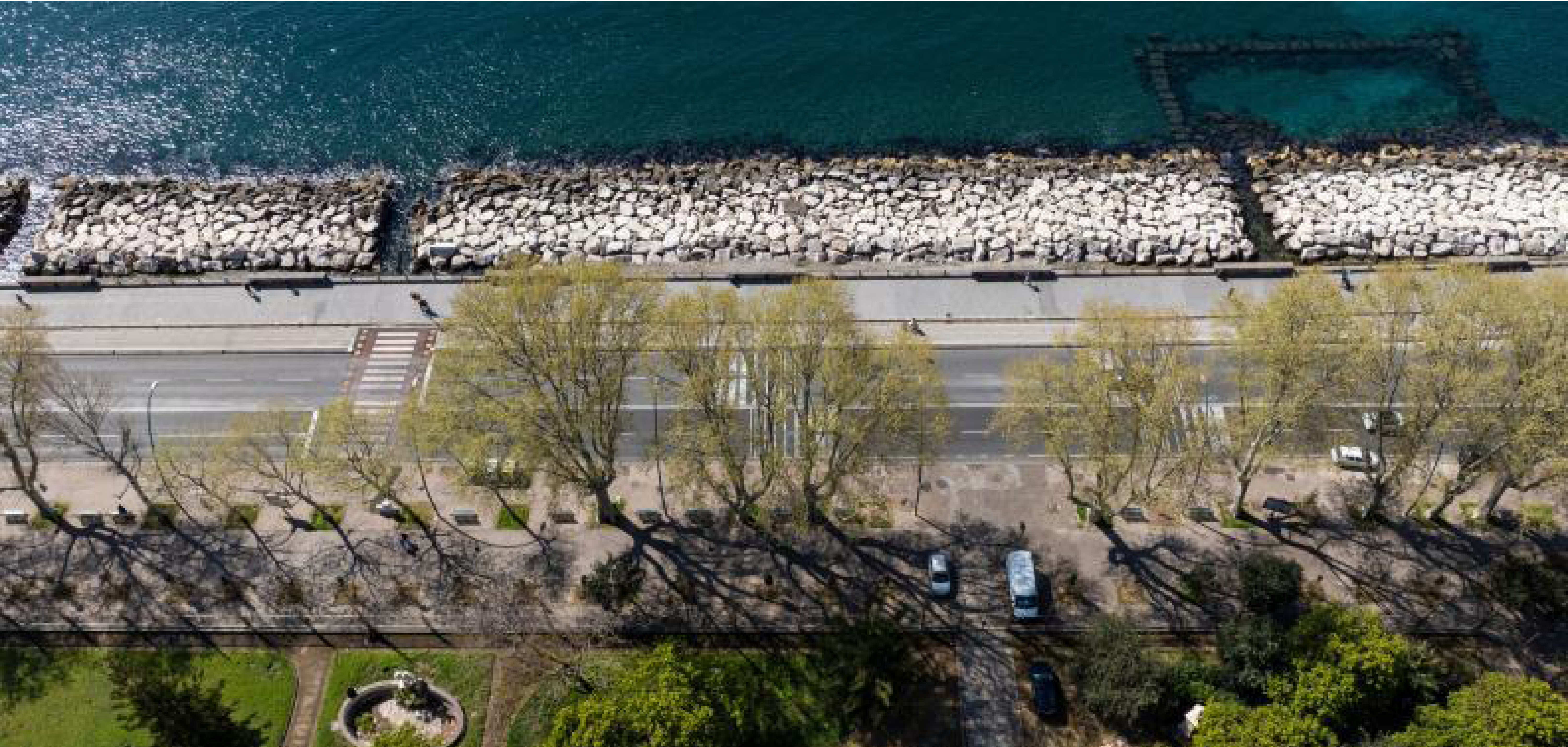
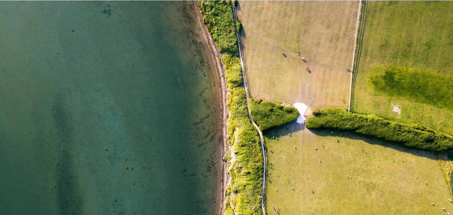
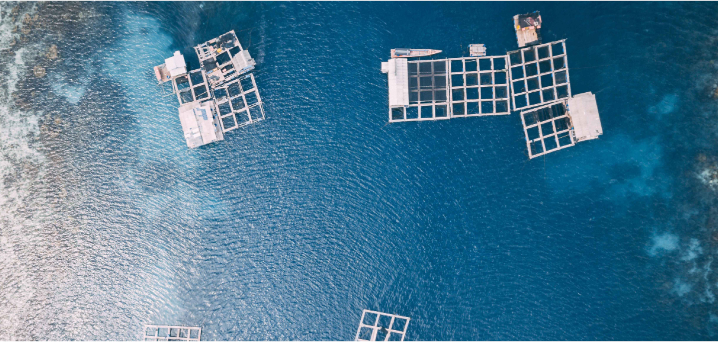
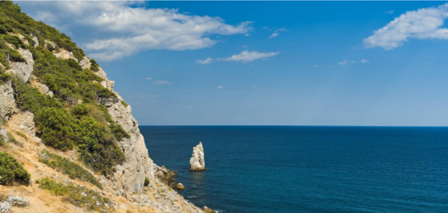
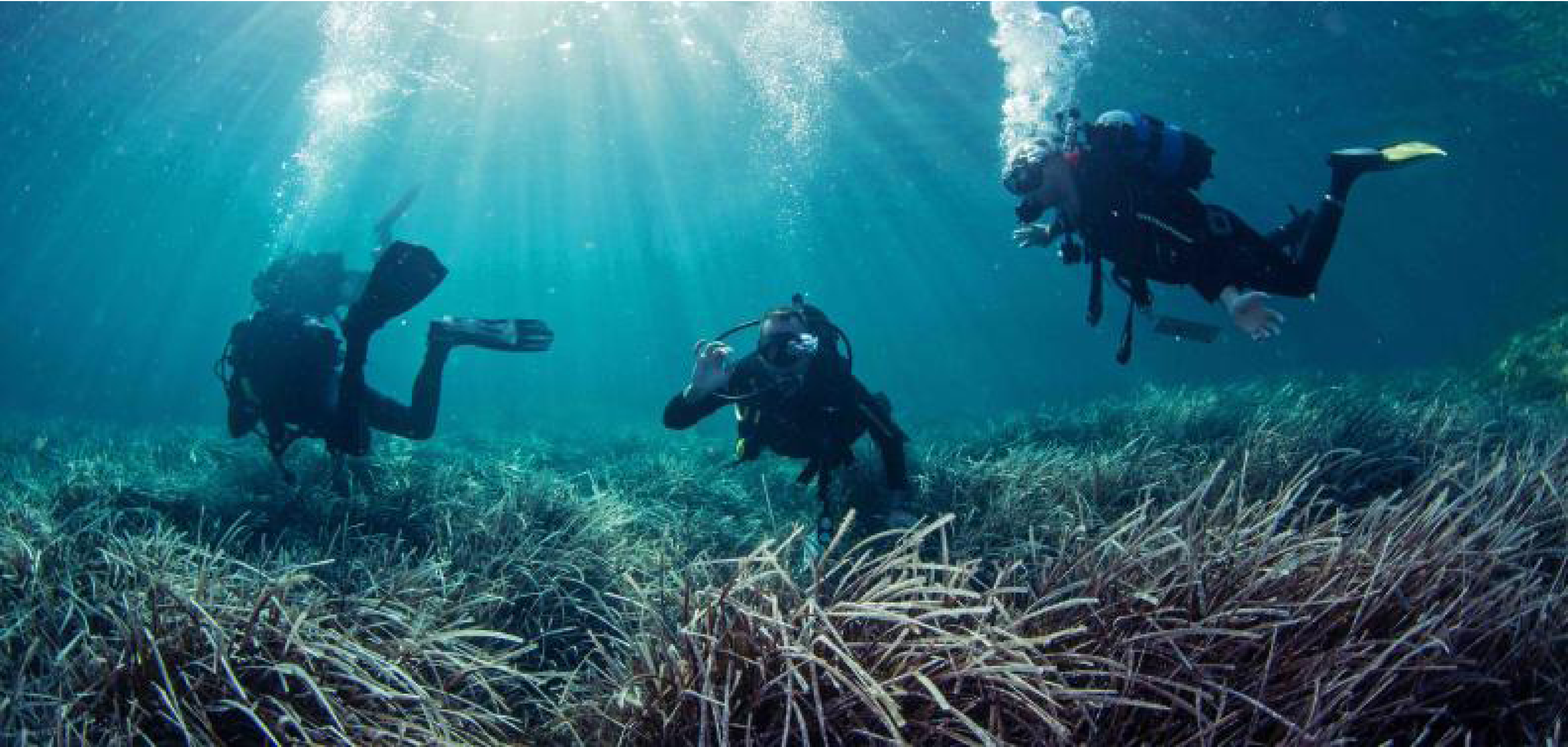
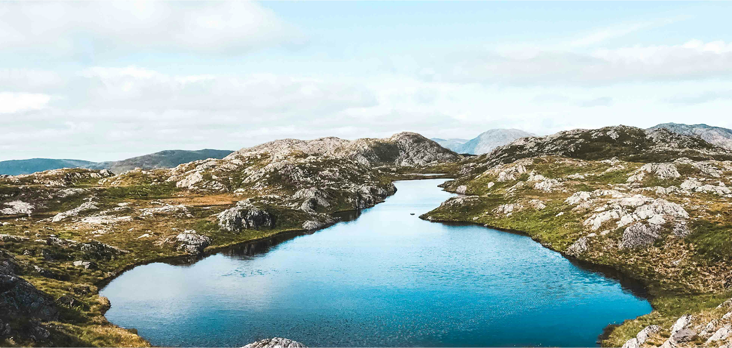
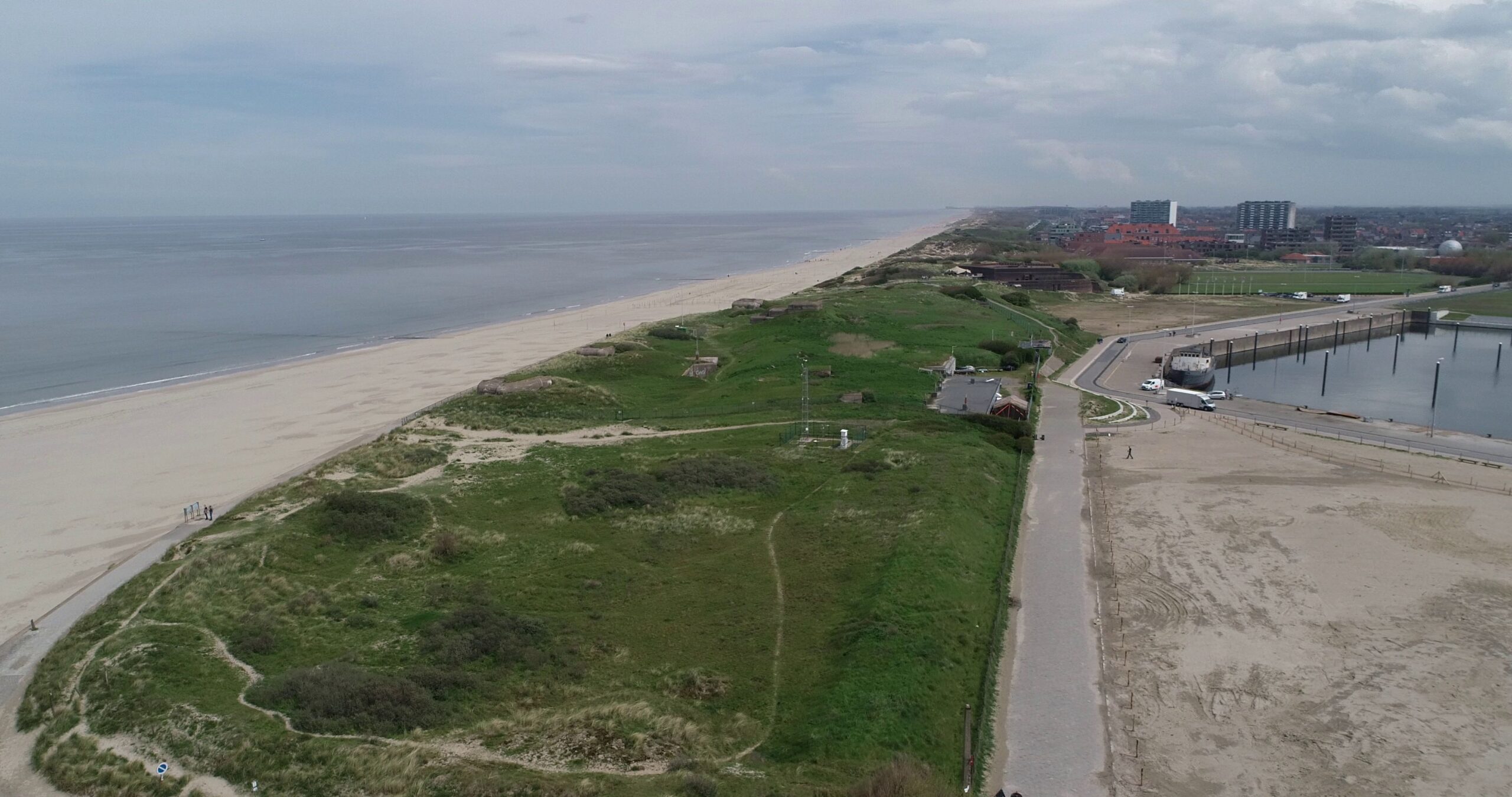
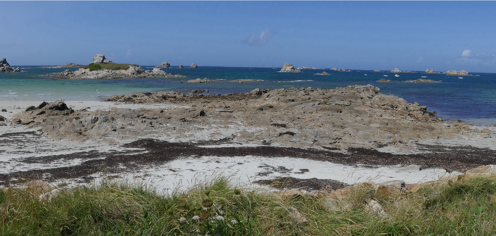
Europe’s coastlines are environments rich in biodiversity that also represent important sites of industry, culture, and heritage. 40 %of Europe’s population live within a coastal region, and many European societies have been, and still are, defined by their relationships with the sea.
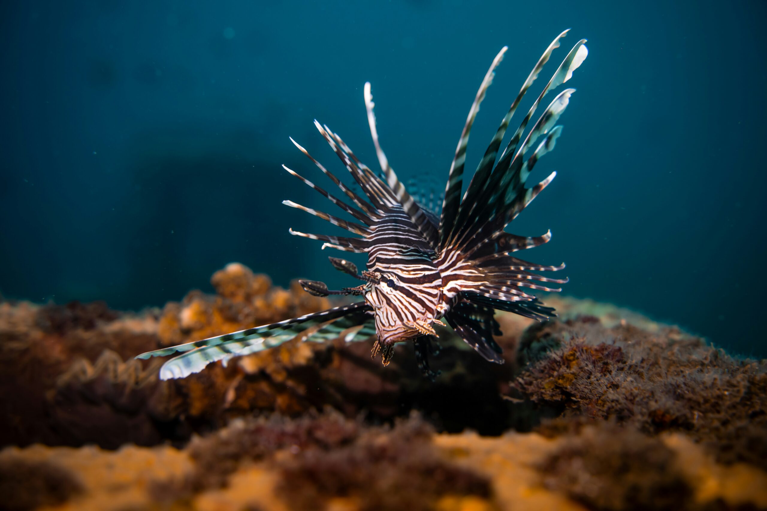
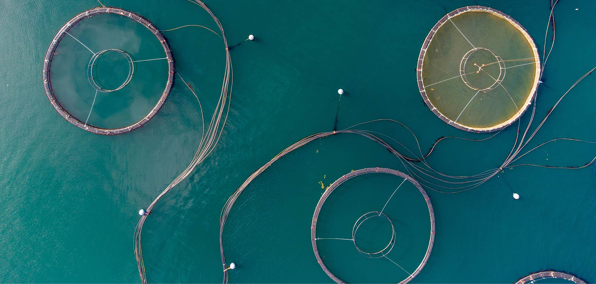
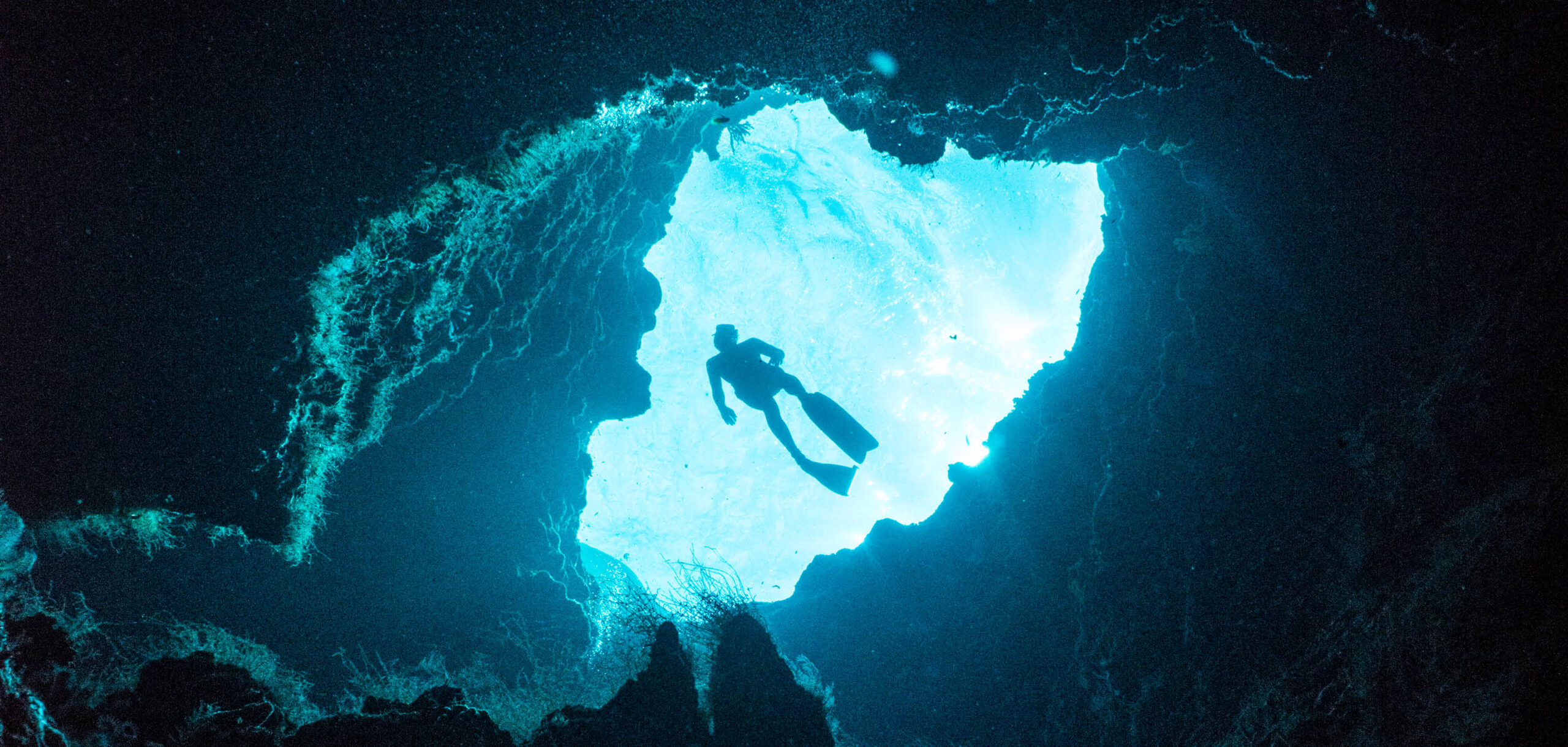
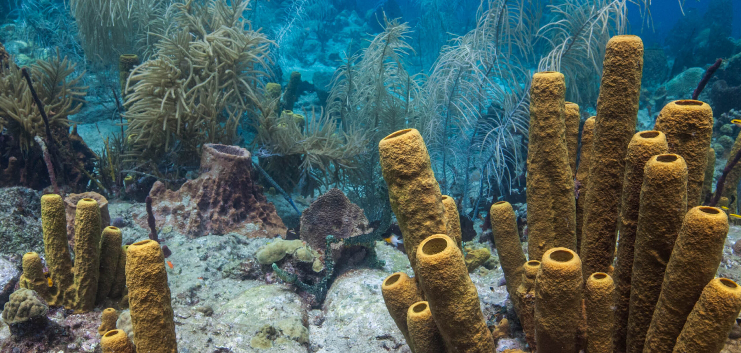
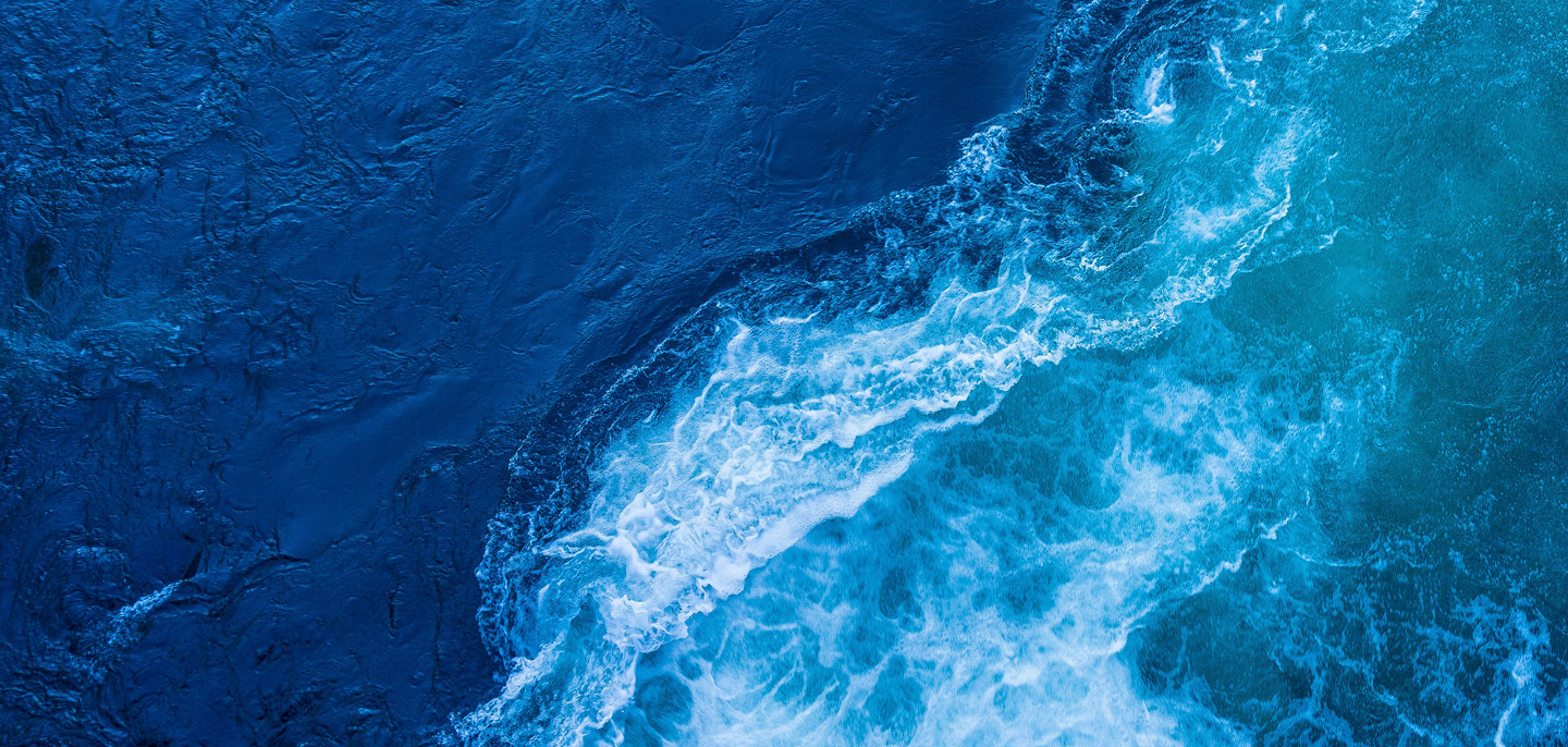
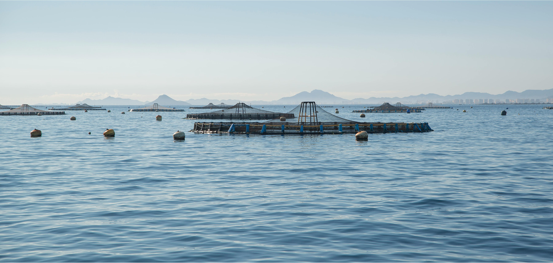
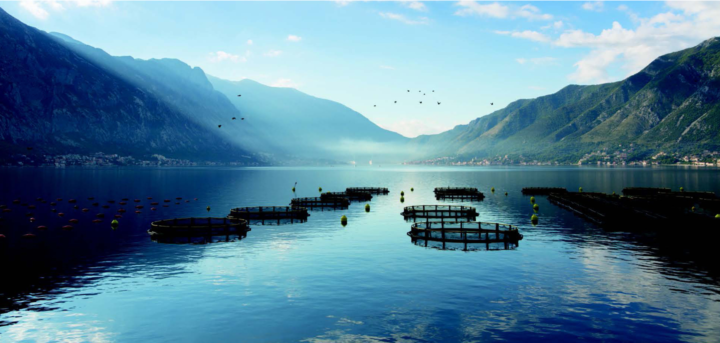
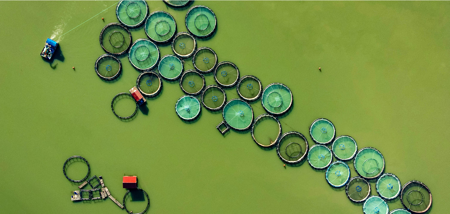
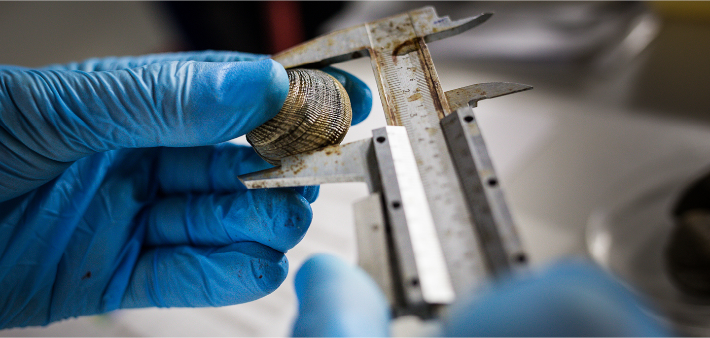
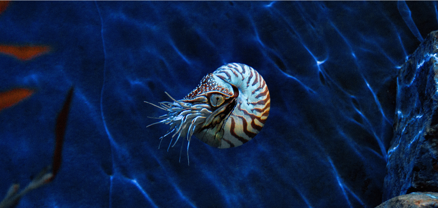
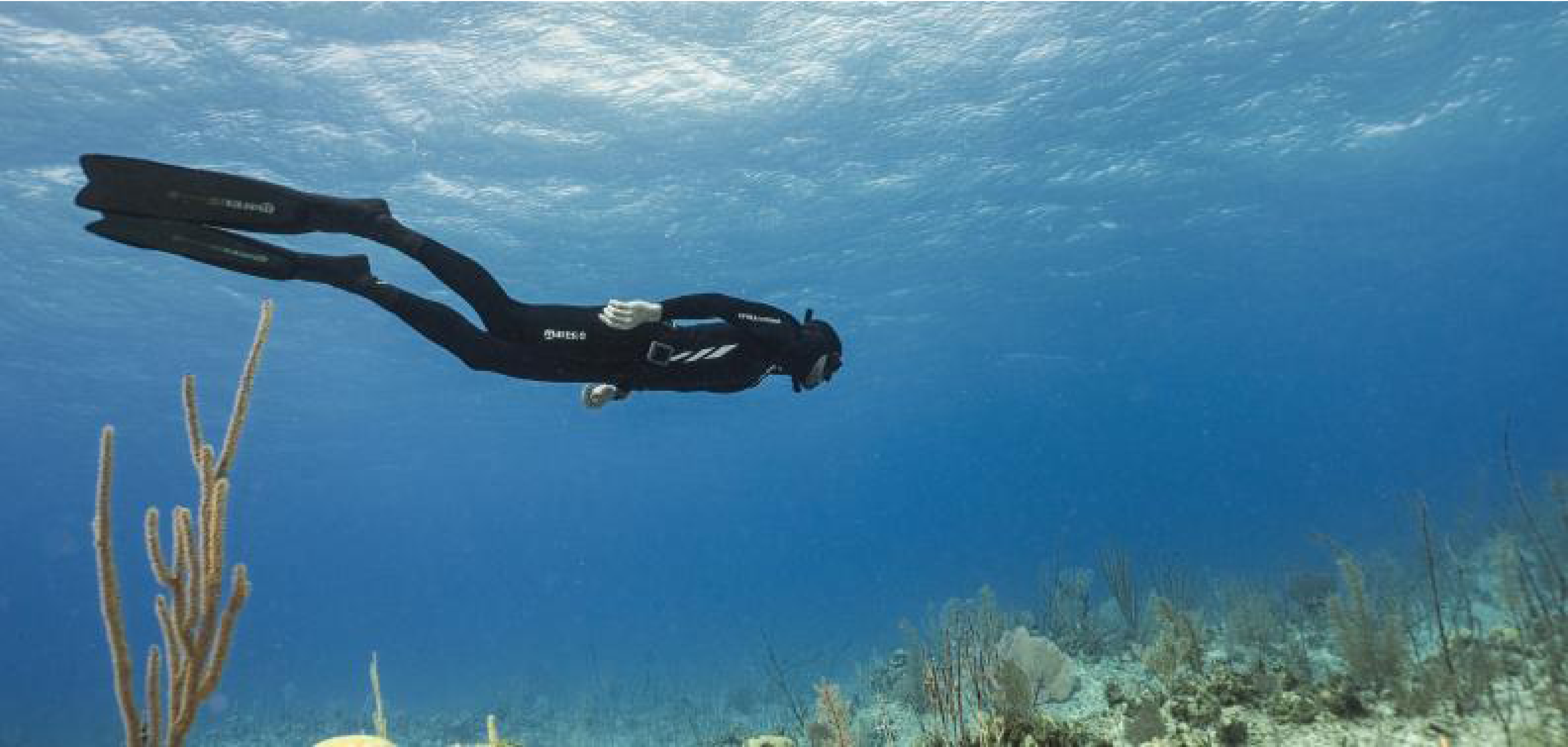
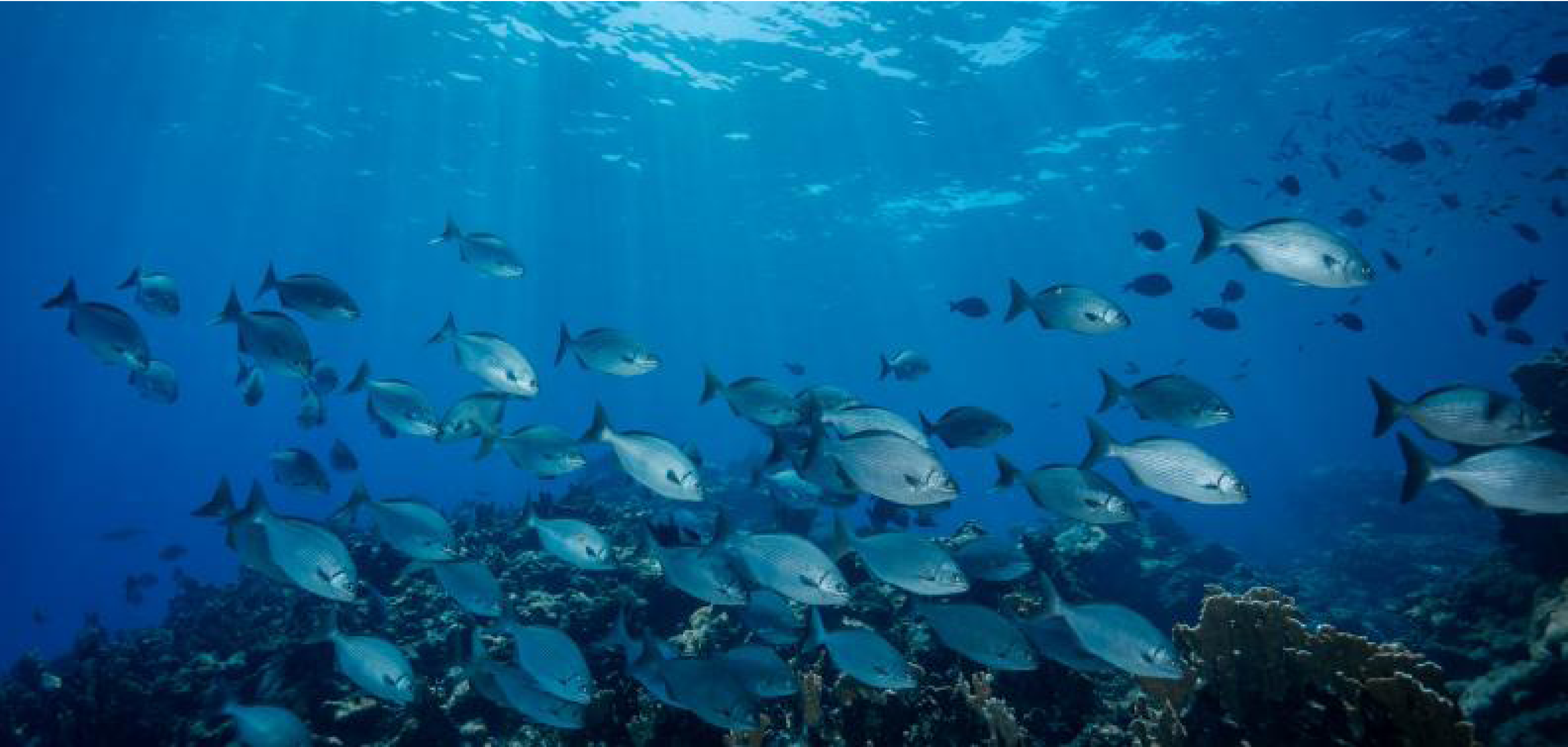
Our seas and coasts represent key ecosystems that host an extremely rich diversity of life and play critical roles in the stability and sustainability of wider ecosystems. However, anthropogenic interferences such as pollution, farming, and building construction, as well as the impact of climate change, are leading to accelerated loss of species’ genetic diversity and destruction of functional ecosystems.
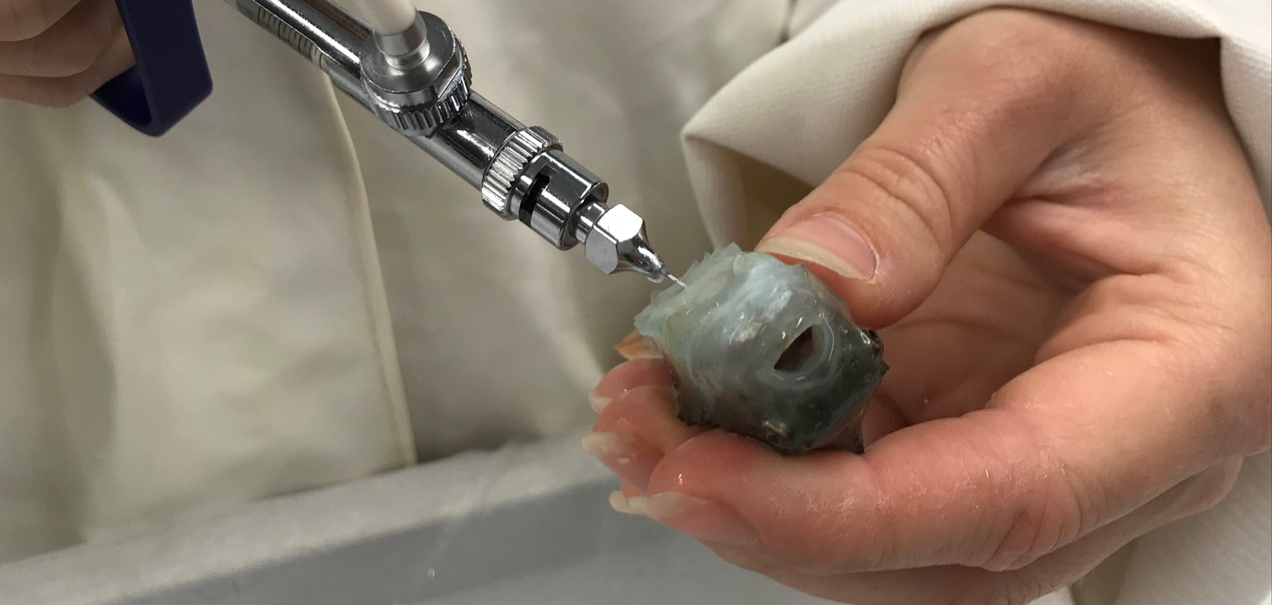
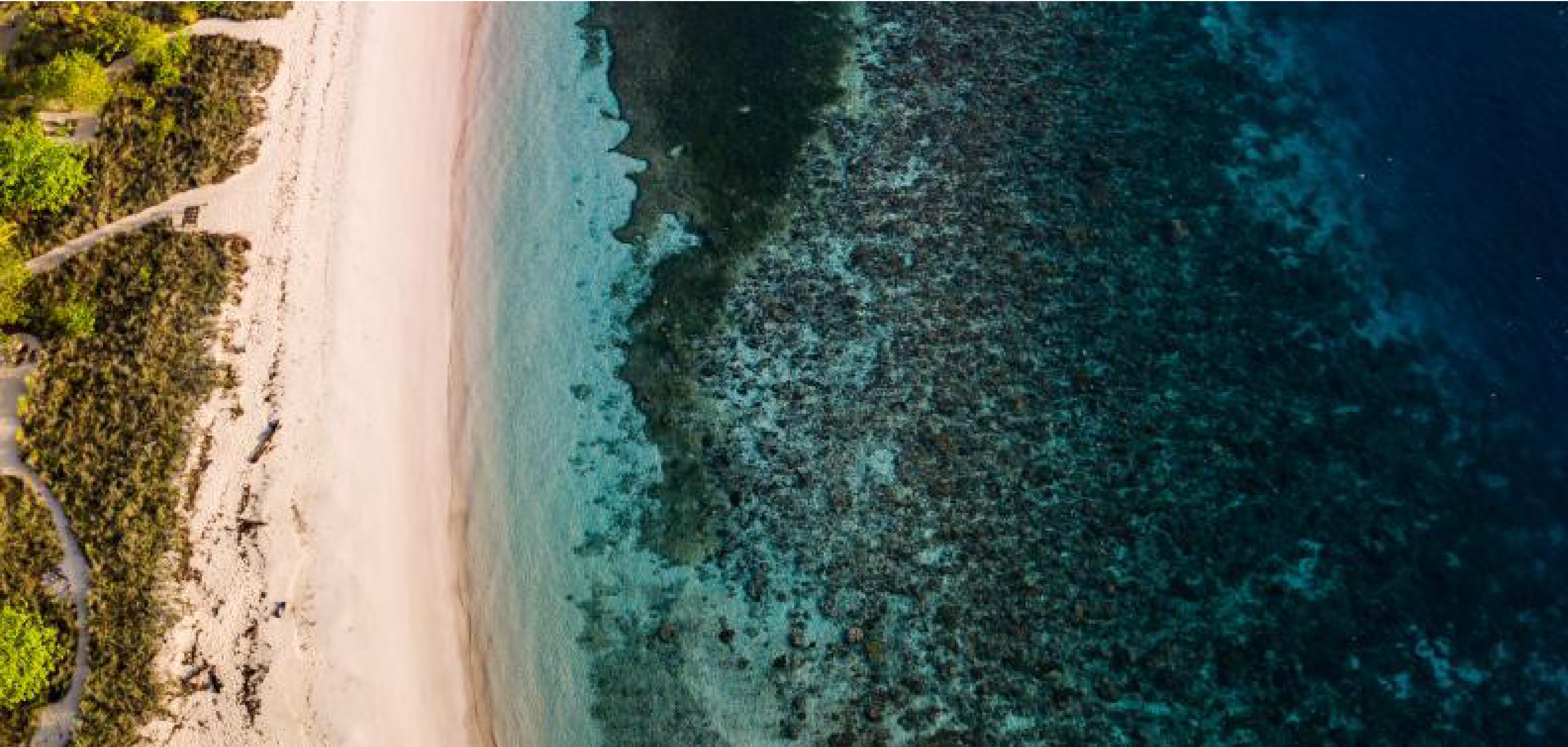
To minimise the future impact of such external factors on coastal biodiversity, we must understand the molecular and cellular basis of how organisms interact in ecosystems and react to external pressures in the context of their natural habitats.
 4 April 2023